Prediction of hemoglobin protein structure and function of the α4.2 deletion mutation and αα/-α4.2/βCD8 (-AA)/βN co-inheritance
DOI:
https://doi.org/10.23917/bioeksperimen.v12i2.16912Keywords:
thalassemia, α4.2 deletion, hemoglobin structure, co-inheritanceAbstract
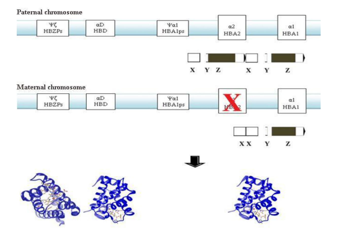
The α4.2 deletion is typically characterized as a silent mutation. The clinical impact of this mutation can vary, particularly when inherited alongside other globin gene mutations, for example co-inheritance with β-thalassemia mutations, such as βCD8 (-AA)/βᴺ. This study aims to determine the α4.2 deletion breakpoint and analyze the structural and functional impact of α-thalassemia heterozygous mutation associated with α4.2 deletion and co-inheritance of αα / -α4.2/ βCD8 (-AA) / βN on the resulting hemoglobin protein. DNA sample was sequenced using the Sanger method by 1st BASE laboratory. Sequence alignment was performed via Clustal Omega, while hemoglobin structure prediction and analysis were conducted using UCSF Chimera (Chimera-1.19) and BIOVIA Discovery Studio. The results identified the α4.2 deletion breakpoint between nucleotide 26 of the X2 box and nucleotide 347 of the X1 box. Despite the mutations, the α4.2 deletion and the αα / -α4.2/ βCD8 (-AA) / βN co-inheritance resulted in a protein structure identical to normal residues, maintaining all stabilizing interactions. Therefore, we conclude that in silico predictions indicate no major structural deviations from wild-type hemoglobin A for both the α4.2 deletion and the co-inheritance of αα/-α4.2 with βCD8 (-AA)/βN. This is consistent with the molecular nature of α-thalassemia deletions, which primarily affect globin gene dosage rather than amino acid sequence, although this observation is limited to computational analysis and has not been experimentally confirmed.
Downloads
References
Ahmed, M. H., Ghatge, M. S., & Safo, M. K. (2020) Hemoglobin: Structure, Function and Allostery. Subcell Biochem. 94:345–382. https://doi.org/10.1007/978-3-030-41769-7_14
Alberts, B., Heald, R., Johnson, A., Morgan, D., Raff, M., Roberts, K., & Walter, P. (2022). Molecular biology of the cell (7th ed., pp. 109–137). W. W. Norton & Company.
Ban, X., Lahiri, P., Dhoble, A. S., Li, D., Gu, Z., Li, C., Cheng, L., Hong, Y., Li, Z., & Kaustubh, B. (2019). Evolutionary Stability of Salt Bridges Hints Its Contribution to Stability of Proteins. Comput Struct Biotechnol J. 17:895–903. https://doi.org/10.1016/j.csbj.2019.06.022
Bhattacharya, R., Rose P. W., Burley, S. K., & Prlić A. (2017). Impact of genetic variation on three dimensional structure and function of proteins. PLoS One, 12(3):e0171355. https://doi.org/10.1371/journal.pone.0171355
Caulier, A., Jankovsky, N., Gautier, E. F., El Nemer, W., Guitton, C., Ouled-Haddou, H., Guillonneau, F., Mayeux, P., Salnot, V., Bruce, J., Picard, V., & Garçon, L. (2022). Red blood cell proteomics reveal remnant protein biosynthesis and folding pathways in PIEZO1-related hereditary xerocytosis. Frontiers in Physiology, 13, 960291. https://doi.org/10.3389/fphys.2022.960291
Dean L. (2005). Chapter 1 Blood and The Cells. In Blood Groups and Red Cell Antigens. National Center for Biotechnology Information. Retrieved January, 25, 2026, from https://www.ncbi.nlm.nih.gov/books/NBK2263/
Handayani, N. S. N., Husna, N., & Sanka, I. (2017). α-globin Alteration in α thalassemia Disorder: Prediction and Interaction Defect. Pakistan Journal of Biological Sciences, 20(7), 343–349. https://doi.org/10.3923/pjbs.2017.343.349
Harewood, J. & Azevedo A. M. (2019). Alpha Thalassemia (Hemoglobin H Disease). In: StatPearls. Treasure Island: StatPearls Publishing. Retrieved January 25, 2026, from https://www.ncbi.nlm.nih.gov/books/NBK441826/
Harteveld, C. L., & Higgs, D. R. (2010). Alpha-thalassaemia. Orphanet journal of rare diseases. 5(13). https://doi.org/10.1186/1750-1172-5-13
He, F. & Jacobson, A. (2015). Nonsense-Mediated mRNA Decay: Degradation of Defective Transcripts Is Only Part of the Story. Annu Rev Genet. 49:339–66. https://doi.org/10.1146/annurev-genet-112414-054639
Hub, J. S., Kubitzki, M. B., & de Groot, B. L. (2010). Spontaneous quaternary and tertiary T-R transitions of human hemoglobin in molecular dynamics simulation. PLoS Comput Biol. 6(5):e1000774. https://doi.org/10.1371/journal.pcbi.1000774
Husna, N. & Handayani, N. S. N. (2021). Molecular and Haematological Characteristics of alpha-Thalassemia Deletions in Yogyakarta Special Region, Indonesia. Rep Biochem Mol Biol. 10(3), 346–353. https://doi.org/10.52547/rbmb.10.3.346
Husna, N., Sanka, I., Arif, A. A., Putri, C., Leonard, E., & Handayani, N. S. N. (2017). Prevalence and Distribution of Thalassemia Trait Screening. J Med Sci. 49(3), 106–113. https://doi.org/10.19106/10.19106/jmedsci004903201702
Keikhaei, B., Bitaraf, S., Mafakher, L., Galehdari, H., Saki-Malehi, A., & Bahadoram, M. (2024). Hematologic parameters cut-off assessment of adult alpha-thalassemia patients in Iran. International Journal of Hematology-Oncology and Stem Cell Research, 18(4), 346–355. https://doi.org/10.18502/ijhoscr.v18i4.16757
Khatri, G., Sahito, A. M., & Ansari, S. A. (2021). Shared molecular basis, diagnosis, and co-inheritance of alpha and beta thalassemia. Blood Research, 56(4), 332–333. https://doi.org/10.5045/br.2021.2021128
Kountouris, P., Lederer, C. W., Fanis, P., Feleki, X., Old, J., & Kleanthous, M. (2014). IthaGenes: an interactive database for haemoglobin variations and epidemiology. PLoS One. 9(7):e103020. https://doi.org.10.1371/journal.pone.0103020
Matthews, K., Lamoureux, E. S., Myrand-Lapierre, M.-E., Duffy, S. P., & Ma, H. (2022). Technologies for measuring red blood cell deformability. Lab on a Chip, 22(7), 1254–1274. https://doi.org/10.1039/D1LC01058A
Nagai, M., Mizusawa, N., Kitagawa, T., & Nagatomo, S. (2018). A role of heme side-chains of human hemoglobin in its function revealed by circular dichroism and resonance Raman spectroscopy. Biophys Rev. 10(2):271–284. https://doi.org/10.1007/s12551-017-0364-5
Nagasurandam, N., Zhu, H., Liu, J., et al. (2015). Analysing the Effect of Mutation on Protein Function and Discovering Potential Inhibitors of CDK4: Molecular Modelling and Dynamics Studies. PLoS One. 10(8):e0133969. https://doi.org/10.1371/journal.pone.0133969
Piel, F. B., Steinberg, M. H., & Rees, D. C. (2017). Sickle cell disease. The New England Journal of Medicine, 384(16), 1561–1573. https://doi.org/10.1056/NEJMra1510865
Sadiq, I.Z., Abubakar, F.S., Usman, H.S., Abdullahi, A.D., Ibrahim, B., Kastayal, B.S., Ibrahim, M., Hassan, H.A. (2024). Thalassemia: Pathophysiology, Diagnosis, and Advances in Treatment. Thalass. Rep. 14, 81–102. https://doi.org/10.3390/thalassrep14040010
Sae-Lee, W., McCafferty, C. L., Verbeke, E. J., Havugimana, P. C., Papoulas, O., McWhite, C. D., Houser, J. R., Vanuytsel, K., Murphy, G. J., Drew, K., Emili, A., Taylor, D. W., & Marcotte, E. M. (2022). The protein organization of a red blood cell. Cell Reports, 40(3), 111103. https://doi.org/10.1016/j.celrep.2022.111103
Singh, V., Baranwal, A. K., Biswas, A. K., Mishra, R. C., & Singh, I. (2025). Prevalence and assessment of the impact of iron-deficiency anemia in beta-thalassemia trait subjects: A study from a tertiary care center of Western India. Asian Journal of Transfusion Science, 19(1), 48–52. https://doi.org/10.4103/ajts.ajts_111_22
Strati, P., Pemmaraju, N., Estrov, Z., Cardenas-Turanzas, M., Pierce, S., Newberry, K. J., Daver, N., Cortes, J., Kantarjian, H., & Verstovsek, S. (2014). Clinical significance of microcytosis in patients with primary myelofibrosis. Leuk Res. 38(10):1212-6. https://doi.org.10.1016/j.leukres.2014.08.007
Taher, A. T., Weatherall, D. J., & Cappellini, M. D. (2018). Thalassaemia. The Lancet, 397(10291), 373–386. https://doi.org/10.1016/S0140-6736(20)32132-0
Viprakasit, V., Ekwattanakit, S., & Riolueang, S. (2022). Clinical classification, screening, and diagnosis for thalassemia. Hematology/Oncology Clinics of North America, 36(2), 193–211. https://doi.org/10.1016/j.hoc.2017.11.006
Wang, Q., He, J., Wu, D., et al. (2015). Interaction of acyperone with human serum albumin: Determination of the binding site by using Discovery Studio and via spectroscopic methods. Journal of Luminescence, 164, 81–85. https://doi.org/10.1016/j.jlumin.2015.03.025
Yang, K., Liu, X., Peng, W., Hua, F., Li, L., Chen, K., Zhang, J., Luo, S., Li, W., Ding, Y., & Xiao, J. (2024). Effects of thalidomide on erythropoiesis and iron homeostasis in transfusion-dependent β-thalassemia. Mediterranean Journal of Hematology and Infectious Diseases, 16(1), e2024001. https://doi.org/10.4084/MJHID.2024.001
Additional Files
Submitted
Accepted
Published
How to Cite
Issue
Section
License
Copyright (c) 2026 Bioeksperimen: Jurnal Penelitian Biologi

This work is licensed under a Creative Commons Attribution-NonCommercial 4.0 International License.